
Veins and arteries of the head and neck

Object numberGC.9173
TitleVeins and arteries of the head and neck
Creator John (1823-1899) Struthers
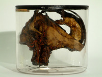
DescriptionThe head and neck of a child, partly cleared of soft parts, and with the veins and arteries injected, the veins painted yellow and the arteries red. The preparation is dried and varnished.
Except for an anterior portion 25 mm. vertically by 30 mm. in transverse diameter the pars frontalis of the frontal bone and the pars orbitalis have been removed. Between paramesial sections 25 mm. part each parietal bone has been removed to the level of the frontal segment. The right zygomatic bone and the right coronoid process of the mandible have been removed and on the left side the posterior half of the mandible. Posteriorly the right half of the occipital bone has been entirely removed as has the left half with the exception of a segment some 20 mm. in vertical depth which retains a connexion with the sagittal segment medially and the bones in the neighbourhood of the asterion laterally. The falx cerebri and the tentorium cerebelli have been retained with the d.ura mater corresponding to the remaining bony portions of the cranium. The proximal five cervical vertebrae are shown and anterior to them the larynx and commencement of the trachea, the hypothyreoid membrane and the deep cervical fascia as it passes from the hyoid bone to. the body of the mandible.
Arteries. The arteries are somewhat eclipsed by the fulness of the injected veins but on the right side the common carotid artery can be observed to its bifurcation lying deep and medial to the external jugular vein. Its bifurcation is obscured but of the external carotid artery there can be distinguished its superior thyreoid branch and lingual artery, the external maxillary artery as it crosses the mandible after giving off its submental branch, and on the face its inferior labial branch is defined. Near the mastoid process the occipital artery crosses the internal jugular vein, gives off its auricular branch and is continued to the cut margin of the occipital bone. Deep to the right external carotid artery can be followed the right internal carotid artery in part of its course, and portions of the right vertebral artery at the level of the atlas are visible anteriorly and again posteriorly as it enters the foramen magnum. On the left side the internal jugular vein is not so full and the left common carotid artery is correspondingly more visible, and its branches, the superior thyreoid, the lingual and the external maxillary, are as clear as on the right side. On both sides the superficial temporal artery is visible in the temporal fossa but more or less overlaid by superficial veins. Only a glimpse is obtained of the left vertebral artery at tie level of the atlanto-epistrophic junction laterally, but within the cranium both vertebral arteries are visible from the foramen magnum to their union as the basilar artery which superiorly divides into its posterior cerebral arteries, from which the posterior communicating arteries go to form the circle of Willis. The posterior communicating arteries join the middle cerebral arteries and thus the anterior cerebral arteries, whose union at the base of the brain by the anterior communicating artery is displayed in this dissection.
Veins. On each side the frontal vein descends on the anterior part of the frontal bone receiving a communicating branch from the ophthalmic vein of the orbit and a communication between the frontal veins where they become the angular veins is effected by branches crossing the nasal bridge. The angular vein descends along the medial and medial half of the inferior margin of the orbit, becoming the anterior facial vein, crosses the maxilla near the angle of which it receives the posterior facial vein, a continuation of the temporal veins and as the common facial vein inferior to the mandible receives a branch from the pterygoid plexus and joins the internal jugular vein about the hyoid level. On either side between the spinous and transverse processes of the cervical vertebrae is a complicated series of veins around a larger vein, the deep cervical vein. On the left side part of the mandible has been removed and the superficial temporal vein with its branches which have been cut short becomes at the level of the zygomatic process the posterior facial vein which, descending, joins the internal jugular vein along with the internal maxillary vein. In relation to the cerebral dura mater the superior sagittal sinus passes along the superior border to the confluens sinuum while the inferior sagittal sinus becoming at the tentorium cerebelli the straight sinus, also passes to the confluens sinuum from which on either side passes the transverse sinus, much larger in this case on the right side than on the left. The course of the right transverse sinus is displayed from/the confluens sinuum to the jugular foremen by removal of much of the occipital bone.
Production periodNineteenth century
Object nameHEAD & NECK
Object categoryAnatomical, specimen
Dimensions
- Container Height: 15.50 cm
Diameter: 17.50 cm