
Lantern slides of WWI field station and notes on wound treatment


Object number@NOO-4
TitleLantern slides of WWI field station and notes on wound treatment
Creator Glasgow RAE Makers
DescriptionPart 4 of 4
Diagrams and graphs of techniques of wound treatment in the field:
Diagram 1 - Cross section of limb to show irregular wound with several perforated tubes distributing hypochlorite solution
Diagram 2 - Dressing with Y connecting tube and two distributing tubes each with four branches so that the whole wound is bathed with the hypochlorite solution
Graph 1 - Microbial chart – Carrel treatment
In upright columns the number of microbes in the microscope field from infinity to 1 and 1 to 1/20: 1/20 means 1 organism found in 20 fields
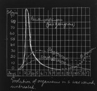
Graph 2 - Evolution of organisms in a war wound, untreated
In gas gangrene, Streptococcus and Staphylococcus
Additional notes written on glass slides are below:
Experience of South African War led surgeons to expect generally aseptic healing of war wounds. After Battle of Marne and Aisne, sepsis found to be invariable and inevitable.
Cause of Sepsis:
1) Composition of projectiles
a. Rifle bullet lead core in casing of cupro-nickel
b. Shrapnel and h.e. (high explosive) ragged pieces of steel
2) High velocity projectiles
3) Infection from ground mud and equipment, gas gangrene, tetanus, streptococci infection
4) Wounded me suffer from hunger, cold, exhaustion, mental excitement.
Development of methods of treatment
Period I: Augt and Sept 1914
Aseptic methods of civilian surgery
Aseptic or antiseptic cleansing of wound suture – results bad
Period II: Oct 1914 to July 1915
Reversion to Listerism
Free opening of wound, drainage, strong antiseptics and antiseptic powders
Pure carbolic
Boisal [??]
Period III: July 1915 to Spring 1916
Physiological method (Almroth Wright)
Hypertonic saline 5% gauze packs or salt tablets –
Non-coagulation of lymph
Increased lymph flow
Bacterial growth prevented
Germs and necrotic tissue washed away – wounds smelly – results good
Period IV: Sept 1916 to autumn 1918
Carrel-Dakin method
• Very free excision of damaged tissue, no suture
• Lavage of wound with hypochlorite of soda 5% (bleaching powder)
• Organisms in wound counted daily
• Sterilisation of wound in 10-15 days
• Secondary suture
Period V: Summer and autumn of 1918
Excision and primary suture (Gray)
• Delayed primary suture
• “Bipp” (Rutherford Morison)
Bismuth Subnitrate – 1 part
Iodofom – 2 parts
Liquid paraffin – q. s.
Wound excised freely, filly with “bipp” and sutured
Phase periods overlapped and the methods practised varied to some degree in different areas with the military exigencies.
‘Discomforts’ of Army Life
• Mud
• Wind – The “Vardar” winds
• Flies
• Mosquitoes
• Extreme cold and heat
• Enemy aeroplanes
• Bombs
• Shells (“Karasouli Kate” and “Lousey Loo”)
Special arrangements in view of British attack on enemy’s position:
1. Preparation of large stocks if dressings, splints etc
2. Arrival of “group” from Base to assist Doctors and Nurses at C.C.S.
a. Group consists of 3 doctors, 5 nurses, 10 orderlies
3. Motor ambulances brought up from base and sent close up to firing line
4. Increase in number of ambulance trains
5. Provisional trains put on disused parts of line (exposed to shell-fire)
Production date 1916 - 1918
Production periodTwentieth century, early
Object namePhotographs, Notes
Object categoryPHOTOGRAPH
Dimensions
- Box Length: 42 cm
Box Width: 12 cm
Box Depth: 12 cm
Slide Height: 8 cm
Slide Width: 8 cm